Dermatological ultrasound · Marrakech

Dermatological Ultrasound in Marrakech: seeing the skin and the tissues beneath it

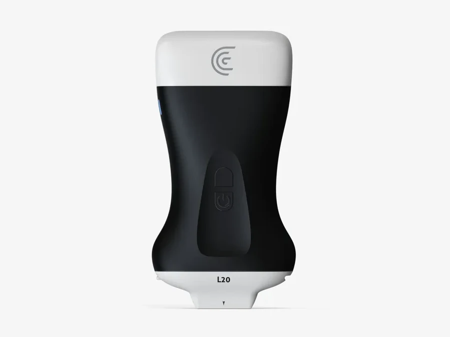
Dermatological ultrasound shows, in real time, the layers of the skin and the tissues just beneath, without radiation and without pain. In Marrakech, Dr Sara Zahid, a dermatologist in Guéliz, has a Clarius L20 HD3 wireless portable ultrasound, fitted with a high-frequency linear probe (8 to 20 MHz) designed for superficial tissues. She performs the examination herself, during the consultation, as a complement to the clinical examination: it helps explore a nodule, cyst or lump, follow certain skin diseases (hidradenitis suppurativa, psoriasis, localised scleroderma) and make aesthetic medicine procedures safer, such as certain hyaluronic acid injections or filler dissolution.

WhatsAppBack to expertise

What is a skin ultrasound?

It is an ultrasound focused on the skin and superficial tissues: epidermis, dermis, fat under the skin, vessels, follicles, nails. A probe placed on the skin, with gel, sends high-frequency ultrasound and reconstructs an image on screen. The higher the frequency, the finer the image of the superficial layers. The examination uses no radiation, needs no injection and takes place during the consultation.

The clinic's device: Clarius L20 HD3

The clinic uses a Clarius wireless portable ultrasound, with the L20 HD3 linear probe (8 to 20 MHz), designed for fine study of the skin. It connects to a tablet or smartphone: the image appears in real time and the dermatologist comments on it with you, at the time she examines your skin.

Exploring a nodule, cyst or lump

When a lesion lies under the skin (nodule, cyst, lump, lymph node, abscess), ultrasound helps specify its size, depth, content and relations to neighbouring structures. It can guide what comes next: monitoring, a procedure, specialist opinion or further tests. It complements the clinical examination and does not remove the need, where necessary, for a biopsy or specialised imaging.

Hidradenitis suppurativa

In hidradenitis suppurativa, an inflammatory disease of the skin folds, ultrasound shows nodules, abscesses and tracts under the skin, sometimes invisible on clinical examination, and specifies their extent. It helps choose treatment, decide on a possible procedure and follow progress under treatment.

Psoriasis and other skin diseases

In psoriasis, ultrasound can help assess skin thickness, nail involvement and, together with a rheumatologist, inflammation of tendons and joints. It also provides information in certain diseases such as localised scleroderma (morphoea), and is used to follow progress under treatment. It is not systematic: the dermatologist decides at the examination.

Aesthetic medicine: locating vessels before an injection

Before certain injections (Botox, hyaluronic acid), notably in vascular-risk areas such as the nose, nasolabial fold or glabella, ultrasound can help locate vessels and their depth, and adapt the technique. It is a safety and precision tool that complements the procedure without replacing it, and without making an injection risk-free: benefits, limits and risks are explained beforehand.

Locating a filler before dissolving it

A lump, asymmetry or swelling after a filler does not always have the same origin: excess product, oedema, inflammation, another cause. Ultrasound shows the filler already injected, its quantity and depth, to better target a possible dissolution with hyaluronidase. It is useful in women and men alike.

How the examination goes

The examination takes place at the consultation. You are made comfortable, gel is applied to the area and the probe is moved gently over the skin. It is painless, without radiation, needs no preparation and the image is commented on straight away. Its duration depends on the area and the question asked. Remember to bring your previous tests (ultrasounds, reports, photos) if you have any.

What ultrasound does not do

Dermatological ultrasound replaces neither the clinical examination, nor a biopsy when necessary, nor specialised imaging (ultrasound of an organ, MRI, CT) ordered from a radiologist. It explores superficial tissues, over a limited depth. The dermatologist always tells you what it lets her see, what it does not allow her to conclude and what comes next.

Where to have a skin ultrasound in Marrakech?

At the Skin & Aesthetic Clinic, in Guéliz, with Dr Sara Zahid, dermatologist, by appointment, in French, English or Arabic. Patients from other Moroccan cities or abroad are also seen, with a first contact possible on WhatsApp.

FAQ

Frequently asked questions

What is a skin ultrasound?

An ultrasound examination that shows in real time the layers of the skin and the tissues just beneath (fat, vessels, lymph nodes, lesions). It is performed by the dermatologist during the consultation, with a high-frequency portable ultrasound.

Is dermatological ultrasound painful?

No. Gel is applied to the skin and the probe is moved gently. An already sensitive area (abscess, inflamed nodule) may sometimes be a little uncomfortable to touch, which the dermatologist takes into account.

Are there rays or risks?

No, ultrasound uses ultrasound waves, not X-rays: there is no radiation. It can be repeated to follow a lesion or treatment.

Is preparation or a prescription needed?

No preparation is needed. The examination is offered at the consultation when useful, without a prescription or prior referral; bring your previous tests if you have any.

Which lesions can be explored by ultrasound?

Mainly lesions under or in the skin: nodules, cysts, lumps, abscesses, lymph nodes, as well as skin thickness and structure. The dermatologist then says what is useful: monitoring, a procedure, specialist opinion, further tests.

Is ultrasound useful in hidradenitis suppurativa?

Yes, it shows nodules, abscesses and tracts under the skin, specifies their extent and is used to follow progress under treatment. It complements the clinical examination.

Is ultrasound useful in psoriasis?

It can help assess the skin, nails and, with a rheumatologist, tendons and joints, and follow progress under treatment. It is not systematic.

Is ultrasound used before or during a filler?

Depending on the indication and area, yes: it allows vessels to be located and a product already injected to be found. It is not systematic, and does not make the procedure risk-free.

Does ultrasound help dissolve a filler?

Yes, it can help locate the product and target the dissolution, notably for a lump or asymmetry whose origin is not obvious. It is not always necessary.

Does ultrasound replace a biopsy or MRI?

No. It complements the clinical examination and can guide what comes next, but replaces neither a biopsy when necessary, nor specialised imaging ordered from a radiologist.

Can men and children benefit?

Yes, the indication depends on the lesion, not on sex or age. The examination is painless and without radiation; the dermatologist decides at the examination whether it is useful.

How much does a dermatological ultrasound cost?

The price is given by the clinic, as it depends on the indication and the consultation. A quote is given before any procedure; there is no single displayed price.

Where can I have a dermatological ultrasound in Marrakech?

At the Skin & Aesthetic Clinic, in Guéliz, with Dr Sara Zahid, dermatologist, by appointment, in French, English or Arabic.

Related conditions

Consult us for one of these conditions

Select your condition: the appointment form fills in automatically.

See all conditions treated
Explore further

A medical & personalised approach

Explore the clinic's complementary expertise or contact us to determine the most suitable care.

Online

👋 Hello! Book an appointment in one click via WhatsApp.

Clinic's professional number